- GU Home
- Paul Ehrlich Foundation
- Early Career Award
- Early Career Award 2023
Prize Winner of the Paul Ehrlich and Ludwig Darmstaedter Early Career Award 2023
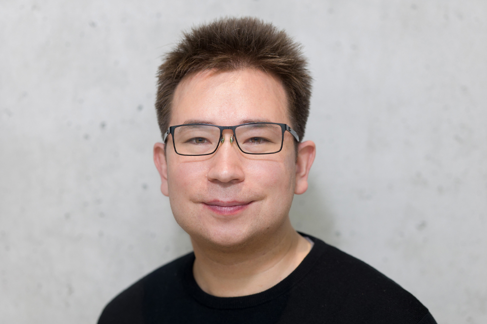

What the mitochondrion tells us
Our blood cells form according to the classical model along four major developmental trajectories: the first trajectory produces the red blood cells, or erythrocytes, that supply us with oxygen; the second delivers the thrombocytes, or platelets, that stop bleeding and enable our wounds to heal. Along the third line, the white blood cells, or lymphocytes, develop that give us our innate immunity, such as the granulocytes, for example, and along the fourth those lymphocytes are produced, such as the B and T cells, without which we would not be able respond to infections with acquired immunity. Even if this categorization is being refined more and more as research progresses, it has provided direction for over a hundred years.
The origin of these developmental trajectories in haematopoiesis (blood formation) are haematopoietic stem cells (HSC), which were discovered a good 60 years ago and isolated for the first time nearly 30 years later. These cells residing in the bone marrow are multipotent, which means that they can transform themselves into variously differentiated daughter cells. At the same time, they are able to renew themselves constantly. Because bone marrow and blood are relatively easy to access, haematopoiesis advanced to become the most thoroughly studied cell development system, and HSC were the preferred model of stem cell research for a long time.
From transplantation experiments to lineage tracing
In the past, it was almost exclusively within animal experiments conducted to test bone marrow transplantation that basic research gained new insights into how the family tree branches out in haematopoiesis. This also resulted in 1961 in the discovery of HSC. Human medicine profited from this through the development of bone marrow transplants, introduced in the 1970s, to treat certain types of leukaemia. Clinical observations led, in turn, to new insights into the processes taking place in haematopoiesis. However, the fact that these insights were obtained under artificial conditions meant that they were limited. After all, the transplanted stem cells and their descendants had been taken from their original context and placed in a new one, where it was possible to trace their developmental potential but not to determine clearly how they behave in natural conditions.
However, thanks to advances in genetic engineering, since the early 1980s a scientific toolkit has been invented in developmental biology and, in decades of work, equipped with increasingly accurate tools that have helped to overcome this limitation in haematological research. It is used to determine both the lineage and differentiation fate of cells in vivo in a process called lineage tracing. To achieve this, genetic markers with fluorescent molecules are implanted in the cells and their transfer from one cell division to the next is traced. These analyses endorse the prominent role of haematopoietic stem cells as sustainable sources for blood formation in healthy organisms. However, they indicate that there are many such sources, from which various developmental pathways evolve that branch out in many different directions. One HSC source might produce only thrombocytes, the other all kinds of blood cells. The stem cell pool is thus heterogeneous, haematopoiesis is polyclonal and the relationships between cells in the blood are unclear. Disentangling them would, however, be important in order to detect, for example, from where a leukaemia cell originates so that it can be better combated, or how to therapeutically apply blood stem cells more effectively. To do this, human medical research must rely on the analysis of naturally occurring markers because, as it goes without saying, lineage tracing with artificial genetic markers in humans is out of the question.
On the track of natural mutations
The most obvious natural markers are somatic DNA mutations, such as occur after each cell division in one daughter cell and not in the other. Such mutations produce a typical genetic pattern that can be assigned, rather like a barcode, to each line of the family tree across cell generations. State-of-the-art sequencing techniques make it possible to hunt for such barcodes throughout the entire genome of bone marrow and blood cells. However, such analyses are expensive, highly error-prone and limited in terms of their informational value. Basing lineage tracing in humans on mutations contained in the genetic information of mitochondria and combining this approach with some of the latest single-cell sequencing technologies, which shed light on the actual health status of the cells under examination, is much more efficient. Leif Ludwig devised this pioneering mix of methods, which is freeing haematology research from its current deadlock, during his time as a postdoctoral researcher at the Broad Institute in Cambridge/USA. As leader of an Emmy Noether Junior Research Group at the Berlin Institute of Health at Charité (BIH) and at the Max Delbrück Center, he is now working on its technical perfection.
Organelles with their own genes
The mitochondria are our cells' powerhouses. From oxygen and hydrogen they produce the energy we need to live. Hundreds to thousands of them are found in most cells, and on average they occupy one fifth of the cells' cytoplasm. They have their own genes, whose information content amounts to around 16,600 base pairs. This genetic text can be checked for changes much more quickly than the 3.2 billion letters of the genetic information in total, especially since many copies of it are present in each cell. This applies even more so given that mitochondrial genes exhibit a mutation rate that is up to 100 times higher than the genes of the cell nucleus. Single-cell sequencing techniques are a particularly reliable method for mapping mutations of mitochondrial DNA (mtDNA).
These single-cell omics, which were voted Breakthrough of the Year by the journal Science in 2018, are based on the separation of complex tissues or mixtures into single cells. Using microfluidics techniques, each of these cells is then packaged in a droplet of liquid and analysed. Three aspects are important here: the messenger RNA pattern of a cell (its transcriptome) shows which of the genes in it have recently been read. Its proteome shows for which proteins these genes have served as blueprints. The open regions of its chromatin, on the other hand, show – like in a live recording – which genes are being laid open at that exact moment for the reading of certain protein blueprints. The DNA threads of each cell nucleus, which are almost two metres long overall, are wound compactly around protein beads in the chromatin. It is only in the temporarily unravelled regions of this tangle that genes can be read. The technique that identifies these areas by labelling them with an enzyme, the transposase Tn5, is called ATAC-Seq.
Two birds with one stone
As mtDNA is not nearly as tightly wound as nuclear DNA, the outcome is that almost all its genes are labelled with Tn5. That is why mtDNA was considered an irksome contaminant in conventional ATAC-Seq protocols. But not by Leif Ludwig. On the contrary: ATAC-Seq enabled him to kill two birds with one stone. From the billions of datasets, it filtered out the mitochondrial DNA data he wanted and at the same time gave him more or less only that information from the nuclear genome that he needed to know in order to identify with which kind of blood cell he was dealing and its current state. In 2019, Ludwig and his team published the proof-of-concept, which showed that the method they had developed for determining cellular relationships (clonal tracking) in the course of haematopoiesis is thousands of times more efficient and also much more informational than methods that sequence the entire genome for this purpose. Just shortly afterwards, they succeeded in extending mitochondrial tracking in vivo to many single cells of different types at the same time. They removed 7,474 haematopoietic stem and progenitor cells from the bone marrow of a healthy 47-year-old donor and three months later 8,591 mononuclear cells from the blood. They used ATAC-Seq to analyse the mtDNA of these cells and assigned them to 12 different cell types on one side and 14 on the other. In this way they identified, for example, multipotent progenitor cells in the bone marrow sample and monocytes in the blood sample. In the process, they found 429 mtDNA mutations that occurred in at least five cells each and discovered 257 different cell families (clones), each with its own mutation profile, which probably originated in 257 different stem cells. Ludwig and his team have also tested this new form of diagnostics on individual patients in a similar way.
Spotlight on the progeny of single stem cells
In total, the number of stem cells that actively contribute to producing around 500 billion blood cells per day is likely to be between 100,000 and 200,000, although there are quite a few on stand-by that only awaken from a kind of slumber if there is an acute need, such as inflammation or blood loss. Essentially, the combination of mtDNA sequencing and single-cell omics devised by Leif Ludwig enables haematology and oncology research and clinical practice to determine for the first time and relatively easily and inexpensively the activity of a single stem cell and the characteristics of its progeny. Such an analysis of the family tree opens up the possibility of finding the branch points where cancer develops or degenerative changes occur during the constant formation of new blood cells and of taking corrective action. In addition, Ludwig's method can be used to study mitochondrially inherited diseases, such as the MELAS syndrome, more comprehensively than before.


CONTACT
Office Paul Ehrlich Foundation:
Friends of the Goethe University Frankfurt
Goethe-Universität Frankfurt
Campus Westend, PA-Gebäude
Theodor-W.-Adorno-Platz 1
60629 Frankfurt am Main
www.vff.uni-frankfurt.de
Managing Directors
Nike von Wersebe
Tel: 069 / 798 12234
Fax: 069 / 798 763 12234
wersebe@vff.uni-frankfurt.de
Julia Lange
Tel: 069 / 798 12452
Fax: 069 / 798 763 12452
E-Mail: lange@vff.uni-frankfurt.de
Adviser for Members & Foundations
Tina Faber
Tel: 069 / 798 17237
Fax: 069 / 798 763 17237
faber@vff.uni-frankfurt.de
Marketing
Constanze von Plato
Tel: 069 / 798 17237
Fax: 069 / 798 763 17237
vonplato@vff.uni-frankfurt.de
Science Communication
Joachim Pietzsch
Tel: 069 36007188
j.pietzsch@wissenswort.com
Bank Account
Paul Ehrlich-Stiftung
Deutsche Bank AG
IBAN: DE38500700100700083900
BIC: DEUTDEFFXXX
Donations are tax deductible.
- Studying at Goethe University
- International applicants
- Faculties
- Overview of study programmes
- Programme for refugees
- GRADE
- Goethe Business School (continuing education)
- Research at Goethe University
- Scientific news
- Goethe Welcome Center (for international researchers)
- Collaborative research projects
- Individual research
- Visiting fellowships
- Endowed chairs
- About the University
- News-in-brief
- University administration
- Campus locations
- Campus life
- University archives (German)
- Rhine-Main-Universities

